Eye Care Services at Good Eye Optometry
We provide routine and medical eye care in a setting built for long-term relationships, diagnostic precision, and clear patient communication.
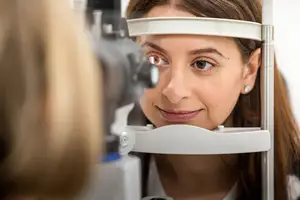
Eye Exams & Medical Eye Disease Management in Brentwood
We care for patients across all stages of life, from first-time eye exams to chronic condition monitoring. Our Brentwood clinic offers comprehensive optometric services, including glasses and contact lens prescriptions, dry eye treatment, myopia management, and medical evaluations for conditions like diabetes and glaucoma risk.
Our approach is diagnostic-first: we evaluate your vision in the context of your overall eye health, visual demands, and long-term needs.
Preventive & Medical Eye Care in One Place
Our services include routine and diagnostic eye exams, prescriptions for glasses or contact lenses, and care for a wide range of medical conditions. We regularly manage dry eye, ocular allergies, diabetic eye health, early signs of glaucoma or cataracts, and pediatric myopia. Patients also come to us with vision changes, digital eye strain, and discomfort that doesn’t resolve with a simple prescription update. Whether you’re here for an annual checkup or ongoing medical care, your visit is paced to allow for clear answers and appropriate follow-up.
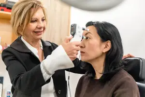
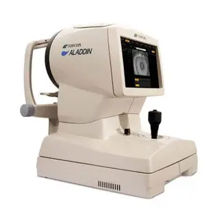
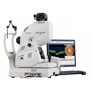
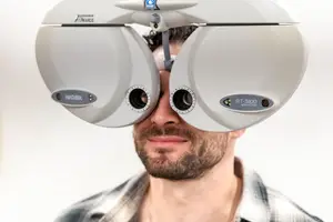
Diagnostic Technology That Supports Better Vision Care
Our Brentwood eye center is equipped with clinical imaging tools that help us assess not just how you’re seeing, but why. As part of our diagnostic care, we use:
OCT (Optical Coherence Tomography) is a non-invasive imaging technology that captures high-resolution cross-sectional images of the retina. At Good Eye Optometry, we use OCT to assess the layers of the retina and monitor the optic nerve for signs of glaucoma, macular degeneration, and other retinal conditions. This test helps us detect subtle structural changes early, often before symptoms appear, allowing for more proactive, informed care.
Meibography and anterior segment imaging allow us to visualize the oil-producing glands in your eyelids (meibomian glands) and assess the front structures of the eye. We use these tools to identify blockages, gland dropout, or surface inflammation that may be contributing to dry eye symptoms.
Corneal topography is a detailed mapping of the cornea’s surface, used to detect irregularities in shape, curvature, and elevation. We use this imaging to guide the selection of contact lenses, especially for patients with astigmatism, keratoconus, or those wearing specialty lenses like ortho-k or sclerals. It also helps us monitor corneal changes over time, ensuring that lens fit remains stable and supportive of long-term eye health.
These technologies help us detect early signs of disease, monitor changes over time, and make sure the care you receive is grounded in accurate, up-to-date data.